Posts

Showing posts with the label Brain

Science is Fun Fridays!

Image
  I came across a book called The Science of Superstition , written by Bruce Hood. A psychologist and philosopher specializing in developmental cognitive neuroscience, he is currently based at the University of Bristol. A major interest has been researching the cognitive processes behind adult magical thinking. In the book, he uses hard science to explain pervasive irrational beliefs and behaviors. Superstitions surrounding Friday the 13th date back to the 19th century, as a pairing, but 13 and Friday have long had ominous connotations. In numerology, 12 is considered complete, while 13 is disruptive.  A 13th guest brings bad luck (Judas in Christianity, Loki in Norse mythology, the devil in ancient Rome). Over 90% of high rises skip the 13th floor due to customer concerns. There's a quote from Swami Vivekananda which says, "If superstition enters, the brain is gone." fMRI has shown that people who engage in superstitious rituals activate the reward centers of the brain, ...

Science is Fun Fridays!

Image
 We have long recognized the importance of music in our lives, throughout history and across cultures. Dr. Oliver Sacks wrote a book about how music can move us, heal us.  People with aphasia, who struggle to speak a coherent sentence, can often still sing - people with Alzheimer's often respond to music even when they respond to little else. Musicophilia Researchers at Anglia Ruskin University have developed a music therapy to assess consciousness levels in children who have suffered significant brain injuries.  MuSICCA, or Music therapy Sensory Instrument for Cognition, Consciousness and Awareness, uses personalized musical stimulation and manipulation to see how the patient responds. A music therapist observes the child's behaviors and records these observations against a set of scales, including responses to various musical stimuli, giving the therapist a picture of the patient's level of consciousness and the strengths and weaknesses of their responses.  The inf...

Science is Fun Fridays!

Image
  An international research consortium has released the first complete wiring diagram of an entire fruit fly brain. Maps that show every neuron in a brain and the myriad connections between them are called connectomes.  Fruit flies have 139,255 neurons in their brain, with over 50 million synapses. That isn't a lot compared to a human brain, which has about 86 million neurons, but fruit flies do complicated things such as walk, fly, navigate, even sing. "If we want to understand how the brain works, we need a mechanistic understanding of how all the neurons fit together and let you think.  For most brains, we have no idea how these networks function," explains Dr. Gregory Jefferis. The FlyWire Consortium brought together expertise from dozens of labs around the world to achieve this.  Dr. Jefferis and other colleagues annotated the different types of neurons found - 4,500 of which were new to science. The CT1 neuron spans across an entire eye and contains 140,000 syn...

Science is Fun Fridays!

Image
  The fusiform gyrus is the part of our brain that processes faces. Some people have face blindness, or prosopagnosia, where the identifying features of a face are difficult to make out.  1 in 50 people experience this, including Brad Pitt and Dr. Oliver Sacks. There's a much rarer condition, called prosopometamorphopsia, of which only 81 cases exist in published literature.  In this, faces are distorted.  One man, Victor Sharrah, recently helped create a computer image of what he sees. "What people don't understand from a picture is that the distorted face is moving, contorting, talking to you, making facial gestures."   The visual can vary - some people see dragon faces or fish heads, and others see horrors like eyes gouged out or cheek cavities. Many people with PMO have been diagnosed as schizophrenic based on presumed hallucinations.  And it's very likely more people have the condition but don't realize they're seeing differently. Catherine Morris...

Science is Fun Fridays!

Image
  With the return of football, let's address CTE, or chronic traumatic encephalopathy.  A recent study shows that early-life concussions are linked with faster cognitive decline. The point being, "even people with traumatic brain injuries in earlier life who appear to have fully recovered from them may still be at increased risk," says author, Marianne Chanti-Ketterl, PhD, MSPH. They used twin participants and found that those injured post age 24 demonstrated pronounced cognitive decline in thinking and memory tests compared to twins without TBI. CTE is more than cognitive impairment though, there are also behavioral and motor symptoms as well.  The damage of repeated concussions can cause the death of nerve cells.  At this time though, it can only be diagnosed upon autopsy. I dislike football, and I honestly think the more we "protected" the players, the rougher they felt capable of being - the aggression has been cultivated.  I also dislike the way it cuts int...

National Memory Day

Image
  Memory is a process of encoding, storing, and retrieving information. Long-term: Episodic memories are the details of past events you have experienced. Semantic memory comprises your knowledge about the world. Procedural memory allows you to learn and execute tasks. Short-term: Working memory is what holds a limited amount of information for immediate mental use.   Sensory memory is our perceptions of the world, and will sometimes persist in long-term memory. Prospective memory is to recall an intention to do something, a more future-oriented memory. The hippocampus and other parts of the medial temporal lobe are critical for many forms of memory, and the amygdala integrates emotional responses into memory. A memory engram is the set of changes in the brain, tracing the synapse connections as they are formed and strengthened. Research has shown that our first memories begin in the womb, but we're unable to recall them.  Most people tend to remember things from abou...

Science is Fun Fridays!

Image
  A distinct layer of tissue has been discovered in the brain, and is one of four such membranes between the skull and the brain. The meningeal layer consists of the dura, arachnoid, and pia mater membranes.  The latest addition divides the subarachnoid space, which contains cerebrospinal fluid (CSF). It is called the subarachnoid lymphatic-like membrane and appears to separate clean CSF from dirty CSF that contains cells' waste products. CSF surrounds the brain and spinal cord, acting as a shock absorber as well as providing immunological protection to the brain.  It also serves a vital function in the regulation of cerebral blood flow. The SLYM was found to be packed with immune cells, which can use the membrane to surveil the CSF for signs of infection or inflammation. Tears in the SLYM can lead to these cells entering the brain, possibly explaining prolonged inflammation following injury.  There may also be implications for Alzheimer's, as a rupture could explain...

Thoughtful Thursday

Image
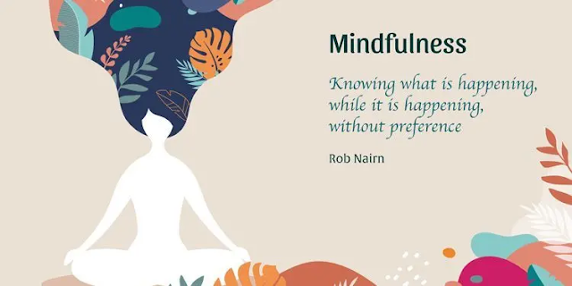
  Definition: A mental state achieved by focusing one's awareness on the present moment, while calmly acknowledging and accepting one's feelings, thoughts, and bodily sensations. I took a college course on Mindfulness from a professor specializing in Acceptance and Commitment Therapy.  He explained thoughts as leaves in the river current, to see them, and to let them float by... I've been practicing a mantra to keep myself centered, to not dwell on my anxieties.  I've come to realize that I've been coping with this for years by making notes, an effort to lessen the weight on my mind. Happy Heart.  This is for today because work has me on edge, but I know I'll get on track. I just need to relax.  :-)

Science is Fun Fridays!

Image
  The "Stoned Ape Theory" suggests magic mushrooms triggered our cognitive evolution. There is no hard evidence for this, but the idea makes sense. There have been studies regarding the mental health benefit of, for example, psilocybin, which activates serotonin receptors and enhances neural plasticity.  Researchers claim that ancient consumption may have allowed for new modes of cognition to emerge, while facilitating brain growth. They further explain that intoxication may have encouraged social bonding through laughter and enhanced the capacity for storytelling and music.  They argue this would have promoted group cohesion and aided in survival. "In particular, the interpersonal and prosocial effects of psilocybin may have mediated the expansion of social bonding mechanisms...imposing a systematic bias on the selective environment that favored selection for prosociality in our lineage." IFL Science

Science is Fun Fridays!

Image
  We are looking at a miniature brain created from stem cells, which have spontaneously sprouted embryonic eye-like structures called optic cups. The cups are two strata of cells which will give rise to the retina.  In this study, they developed several types of retinal cells that organized into neuronal networks and displayed connectivity with other regions of the brain organoid, becoming fully integrated and responding to light.  This allowed researchers a glimpse into the interactions between the eyes and brain of a developing embryo. Researchers were able to reproduce these findings - out of 314 mini-brains, 72% grew the optic cups. "These organoids can help to study brain-eye interactions during embryo development, model congenital retinal disorders, and generate patient-specific retinal cell types for personalized drug testing and transplantation therapies." Study author, Jay Gopalakrishnan IFLScience

Science is Fun Fridays!

Image
  A recent experiment studied people learning to use a specially designed robotic "third thumb."  Not only did they manage with ease, but their brains quickly adapted to the new skill as well. The thumb is controlled using a wireless pressure sensor attached to the underside of the foot.  The user applies a small amount of pressure under their big toe and the thumb contracts for grabbing. 20 participants were trained to use the thumb and then encouraged to use it at home to complete tasks, totaling 2-6 hours per day.  Another 10 participants wore a static thumb. Using an fMRI scanner, they asked them to move their fingers individually, and found a small but significant change to the way hand movement is represented in the sensorimotor cortex in those who had used the robotic thumb.  The changes did not persist once the participants stopped using the extra thumb, but the findings demonstrate our brain's amazing plasticity and capability to adapt to technological ...

Science is Fun Fridays!

Image
Researchers have found that in the hours after we die, some brain cells not only remain active, but increase their activity. These "zombie genes" are glial cells, which will grow and sprout appendages, but that in itself isn't too surprising.  These cells are responsible for clean up after brain injuries such as oxygen deprivation and stroke. However, not knowing this before may complicate some of the research done using post-mortem brain tissue to find treatments and cures for such diseases as Alzheimer's and schizophrenia, because it has not accounted for the post-mortem cell activity. "Most studies assume that everything in the brain stops when the heart stops beating, but this is not so." -Dr. Jeffrey Loeb, head of neurology and rehabilitation at UIC College of Medicine. Loeb and his team noticed that the global pattern of gene expression in fresh human brain tissue didn't match any of the published reports of post-mortem gene expression, so they dec...

Science is Fun Fridays!

Image
  With regards to neurodegenerative diseases such as dementia, scientists believe that disrupted protein synthesis is a major hallmark of aging.  Through wear and tear they exhibit inflammation and infection, which can impair the protein production that mediates cellular function.  To prevent these cells from causing harm, an "integrated stress response" becomes activated which shuts down protein production entirely. They believe as we age, the ISR may become too active, so they wanted to test if it could be inhibited and thus reboot protein production. Aged mice were trained to escape from a water maze using a hidden platform.  Using spatial, working, and episodic memory makes this harder for older mice to complete.  When the mice were given the ISR inhibitor over a period of three days, their performance levels matched those of younger mice. They were tested again in a constantly changing maze, and they again found that the older mice treated with the inhibito...

Science is Fun Fridays!

Image
  In 79 AD, Mount Vesuvius erupted, burying the towns of Pompeii and Herculaneum.   A cloud of scorching ash buried a young man in Herculaneum.  As the ash cooled, it turned some of his brain to glass.  Above, we're looking at some of his nerve cells. Pier Pablo Petrone is a biologist and forensic anthropologist at the University of Naples Federico II in Italy.  He has found brain samples before, but says they were of a soaplike substance or were mummified. But in this case, the brain turned to glass as it was heated, liquefied and cooled.  This process is called vitrification.  Petrone and colleagues used electron microscopy to study the remains - they found layers of tissue wrapped around tendrils in the brain tissue and concluded it was myelin, a fatty substance that helps carry signals along nerve fibers. "This is the first ever discovery of ancient human brain remains vitrified by hot ash during a volcanic eruption." Science News

Science is Fun Fridays!

Image
  Comparative neuroanatomy searches for the rules behind such organized diversity, as seen above.  All of these brains have a cerebral cortex, a cerebellum in the back, olfactory bulbs up front, and a brainstem that is connected to the spinal cord. The marmoset brain stands out a little, at least in my opinion.  It is the most smooth, with almost no visible gyri (folds) or sulci (grooves) on the surface.  This makes the marmoset brain popular for studies of the visual system - the vast majority of the visual cortex lies exposed. Comparative neuroanatomy is used by paleoneurologists to understand the evolution of primate and human brains, to assess the nature of our cognitive abilities.  Herculano-Houzel notes that it's less about gross anatomy, and more to do with architecture and neuronal connections.  She found that the horizontal spacing distance between neurons have increased in humans in the frontopolar cortex as well as what we call Broca's area, whic...

Science is Fun Fridays!

Image
Researchers at Tel Aviv University have conducted a study looking at brain connectivity in 130 mammalian species, including humans. The results contradict years of assumptions, but shouldn't really be that surprising.  "We discovered that brain connectivity - namely the efficiency of information transfer through the neural network - does not depend on either size or structure of any specific brain." "The brains of all mammals, from tiny mice through humans to large bulls and dolphins, exhibit equal connectivity." The brains were from already dead animals obtained from the Kimron Veterinary Institute. Many of these animals' brains had never before been MRI scanned, and so a unique database has also been generated through this study. The technology used detects white matter in the brain, and enabled researchers to reconstruct a neural network - neurons and their axons through which information travels, and the synapses where they meet....

Science is Fun Fridays!

Image
Mouse Brain Splices Mice have 100 million brain cells, whereas humans have about 100 billion.  But for years, scientists have turned to mice for study, because the cell types are similar in the cortex. Neuroscientists at the Allen Institute for Brain Science have created a high resolution 3D atlas of a mouse brain. In 2016 they released a 3D mapping of the cortex, as part of the Mouse Common Coordinate Framework (CCF). And now, CCFv3 can pinpoint individual cells' location.  The construction came from the average brain anatomy of almost 1,700 mice. SciNews Allen Institute

Book Club - February

Image
In a college course, a professor read excerpts from Oliver Sacks' The Man Who Mistook His Wife for a Hat.   After class, I sought the book out for myself, and was mesmerized by the case studies he shared.  It also helps that Sacks is such a storyteller - it doesn't feel clinical when he talks about these medical issues.  I was already interested in the brain and cognition through my psychology courses, but Sacks really influenced my interest in brain injuries and neurological disorders. From there I wanted to read all his works - to date, I've read Musicophilia, Hallucinations, and Awakenings.  I've read portions of The Mind's Eye , so that's technically on my list still. This month, I intend to start An Anthropologist on Mars. I know one of the essays is about Temple Grandin, an autistic woman who designed humane livestock facilities, inspired by a "hug machine" she had built for herself.  The effect is that deep pressure is calming ...

Science is Fun Fridays!

Image
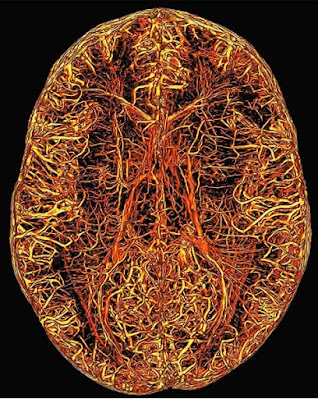
Deep Vascular Architecture The brain fascinates me in so many ways, it's such an amazing organ both in function and form.  I found an Instagram page called 'Neuroradiology' and I have been loving the images. Diffusion tractography Brain vessels It also introduced me to a neuroscience artist, Greg Dunn. Cerebellum and brain stem. Microetching Neuroradiology Greg Dunn